|
||||||
|
Dermatoscopy with digital epiluminescence
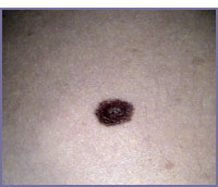
Dermatoscopy with epiluminescence is a non-invasive diagnostic method, which allows to evaluate the pigmented cutaneous lesions, eliminating the incidental visible light radiation and recording the image with a digital video-camera. With this method it is possible to recognize the disposition of pigment in all levels of the epidermis (intraepidermal, Junctional and dermal), in fact pigmentation has different morphologic characteristics in the various melanocytic lesions of the skin.
The digital record of the images allows a precise follow-up of the lesions and the evaluation of eventual modifications in the disposition of pigment, which give a precocious diagnosis of an initial malignant transformation. The different characteristics are: 1) Colour: the depth of the pigment causes variation of colour, in fact melanin in corneum is black, in lower epidermis and junction is brown, grey in dermis and blue in deep dermis. 2) Pattern of pigmentation: normally the pigmentation forms a reticulum (reticular pattern), characterized by a net of pigmented lines, corresponding to the pigmented basal layer. The features of reticulum are important criteria for diagnosis, in fact irregularity of reticulum is found in “atypical moles”. Another pattern is the “globular”, characterized by pigmented spherical bodies, which correspond to nests of nevic cells at the dermo-epidermal junction; as in the reticular P. the regularity of globules is a favourable criterium. Another Pattern is the “Diffuse”, due to large amount of pigment in all cutaneous levels, and can hide the reticular Pattern. An irregular Diffuse pattern and the presence of a “grey.blue veil” is diagnostic for an atypical mole versus melanoma. The “Ivy-leaf areas”, are zones of irregular pigmentation at the periphery of a basal cell carcinoma. The De pigmentation, defined as “Regression” is a reduction of pigmentation inside a pigmented lesion, normally It is a physiologic event in the life of a nevus, but rarely can be interpreted as a malignant transformation. 3) Pseudopodes or radial striae: are periferical estensions of pigmentation, typical of Spitz Nevus and rarely a radial-growth of a melanoma 4) Corneum pseudo-cysts and comedo-like ducts are typical of seborrheic keratosis. |