|
||||||
|
Histologic and cytologic examinations
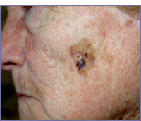
Histologic and cytologic examinations: in cutaneous disease, histologic and cytologic examinations are very important, in order to permit a precise diagnosis and a subsequent correct therapy.
The cytologic examination consists in taking a scraped sample of the skin, with a sterilized scalpel. The scraped material is put on the glass slide, then coloured with Papanicolau’s method or with Giemsa stain and observed with microscope. This method points out the nuclei of the cells, allowing to recognize neoplastic from normal cells, cells infected by viruses and characteristic epithelial cells of particular cutaneous diseases (acantholitic diseases). The presence of neoplastic cells gives the choice of a correct therapy in neoplastic diseases: between a surgical approach in aggressive cutaneous timors and physical therapy (cryotherapy, diatermocoagulation) in precancerous lesions, in this case sparing surgical procedures. The histologic examination is made on a surgically excised sample of cutaneous tissue: a very thin section of the tissue is cut and then coloured with specific methods; the microscopic vision of the cytoarchitecture of the lesion allows a more precise diagnosis. |