|
||||||
|
Pigmented lesions of the skin
Melanocytic Junctional Nevus: it is a proliferation of melanocytes at the dermo-epidermal junction. Clinically is a flat pigmented lesion (“mole”) with omogeneous colour, generally with symmetric shape (typical nevus). It is a benign lesion. Melanocytic Composed Nevus: it is a proliferation of melanocytes at the dermo-epidermal junction and in the dermis. It is considered a maturation of a junctional nevus. Clinically it is a papulo-nodular pigmented lesion (“mole”) with omogeneous colour and with symmetrical shape (typical nevus). It is a benign lesion. Melanocytic dermal nevus: it is the last phase of the natural life of a nevus, with proliferation of melanocytes only in the dermis, which clinically corresponds to a dome-shaped lesion, generally pigmented. It is a benign lesion.
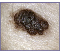
Blue nevus: it is a proliferation of particular spindle-shaped melanocytic cell in the dermis. This deep proliferation gives a blue coloration to the skin. Clinically it is a dome-shaped lesion with a dark-blue colour. It is a benign lesion. Spitz Nevus: it is a benign nevus, which can simulate a melanoma. Clinically it is a rapidly growing symmetric nodular lesion, pigmented or not pigmented (red-colour), which appears generally in young people, on all areas of the skin . Generally it is surgically removed. The dermatoscopy can show specific features, sparing a surgical incision. Melanoma: is the malignant tumour of melanocytic cell. Clinically It is a growing pigmented lesion, arising de novo in the epidermis, or in a pre-existent melanocytic lesion, with irregular chromatic and morphologic features. It usually appears around the age 40-50 years, but recently it has been found in younger adults; before adolescence it is an extremely rare event. |